


LSU Research Bites: New 3D Tool Helps Prevent Joint Damage in Horses
April 13, 2026
For horses—and for humans—joint injuries and chronic joint stress can lead to degenerative joint disease. The way we deal with this often involves medications that target pain or inflammation.
Historically, we’ve failed to address the underlying cause of the original injury, such as problematic gait patterns or what are called “pathomechanical” forces.
A more whole-body approach to addressing and preventing injury is thankfully becoming more common among human athletes but remains rare in equine care and research.
Michelle Osborn, an associate professor in the Department of Comparative Biomedical Sciences at the LSU School of Veterinary Medicine, is working to change the paradigm.
“Our long-term goal is to establish pathomechanical models of equine lameness that can be used to develop individualized preventative and rehabilitative therapies,” Osborn said. “To do this, we created a realistic, dynamic 3D equine skeletal model that is capable of accurately modeling the movements of horses.”
Osborn and colleagues recently published a case study in Frontiers in Veterinary Science of a 3D equine skeletal model, a tool that can be used to analyze posture and movement and objectively document the effectiveness of training programs in changing movement patterns.
“Designing targeted therapies for horses requires recognizing habitual postures and movements that are pathological,” the research team writes in the case study.
The model, built from CT imaging data of a horse, can animate working postures before and after a whole-body exercise regimen to illustrate how interventions can correct harmful movement patterns and the forces they place on specific joints.
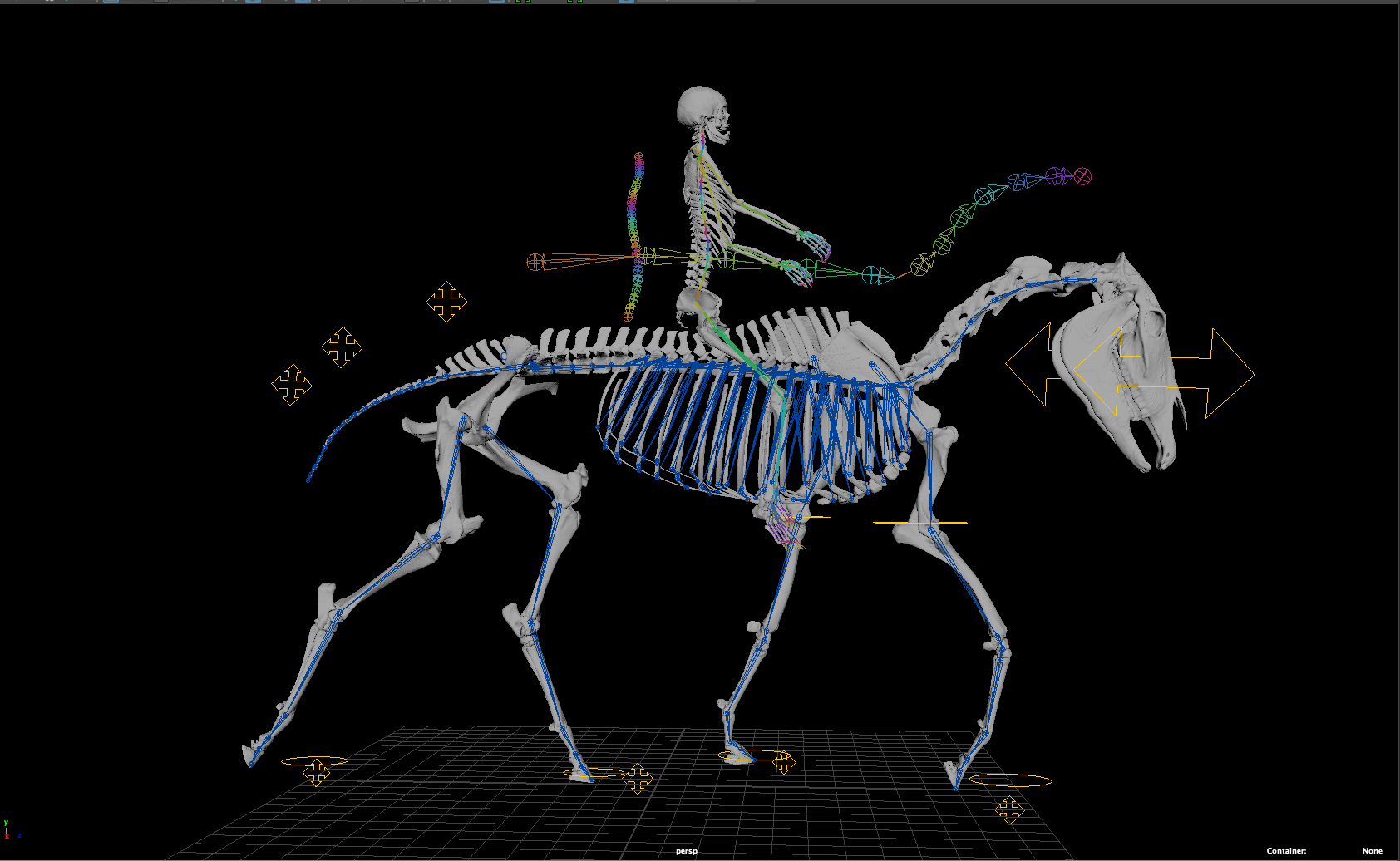
The horse-and-rider combined model offers a way to visualize movement in areas of the horse and human bodies that are otherwise difficult to see, such as within their spines.
“CT scanning of an entire horse is a rarity, but technology is quickly improving and, perhaps, will one day be more commonplace,” Osborn said. “Until that happens, our model can be adjusted in size to better fit a horse as depicted in videos or photographs.”
A horse's joint angles during movement, which can be analyzed using freeze-frame images from a video, for example, are currently hand-matched to skeletal elements of the 3D model and tweaked in model animations to demonstrate how movements are transferring forces through the horse’s joints.
“We are currently working to automate this process so that the movement of the horse can be directly linked to movement on videos using visible joint angles or, when available, surface markers,” Osborn said.
“While motion capture data is always best for scientific purposes, we want our model to be as useful for riders and trainers who are working with their horses in the field as it is for scientists with experimental setups.”
The research team is now working to optimize their model for eventual release to horse riders and trainers.
But case study reviewers have already expressed interest in the whole-body exercise therapy used in the case study, based on the 3D model’s demonstration of its beneficial effects on joint movement and forces.
“The role of mechanics in the formation and function of tissues in health and disease is only just beginning to be recognized as being just as important, if not more so, than chemistry and genetics,” Osborn said.
Research is finding that mechanical overloading of joints can actually change gene expression and induce the release of inflammatory mediators that cause inflammation. The effects are separate from the actual tissue damage itself caused by joint overloading.
“This explains why the common use of anti-inflammatories as treatment, while allowing mechanical dysfunction to continue, results in having to increase the dose given, and also why they eventually fail to control pain,” Osborn said.

“We want our model to be as useful for riders and trainers who are working with their horses in the field as it is for scientists with experimental setups.”
Michelle Osborn, associate professor, LSU School of Veterinary Medicine
The team is excited about the potential of the 3D model to serve as a tool that can directly benefit the lives of horses and riders. The horse skeleton model, a tool for modeling whole-body posture, movement, and analysis of 3D force regimes, can also be combined with a dynamic 3D human model to understand the relationship between horse and rider.
The horse-and-rider combined model offers a way to visualize movement in areas of the horse and human bodies that are otherwise difficult to see, such as within their spines.
Osborn started this line of research as a postdoctoral researcher at the University of Georgia School of Veterinary Medicine.
There, she worked with veterinary pathologist Dr. Betsy Uhl and her horse trainer and equine expert Jean Luc Cornille to better understand the complex relationship among posture, movement, forces, and tissue degeneration, with the goal of providing information that is useful and practical for those who work with horses.
At LSU, Osborn has continued her collaboration with these experts and has also involved LSU graduate and undergraduate students in the research.
“Our work to improve the model and use it to depict more cases not only strengthens our interinstitutional collaboration, but it also continues to provide rich opportunities for undergraduate, graduate, and professional student researchers,” Osborn said.
Read the case study: Frontiers | The dynamic 3D horse: analyzing the relationship between whole body pathomechanics and joint degeneration in the fetlocks